Dataset for Optical characterization of magnesium incorporation in p-GaN layers for core–shell nanorod light-emitting diodes
This dataset contains the results of scanning electron microscopy (SEM), micro-photoluminescence (PL), cathodoluminescence (CL), Raman, and Electron Beam Induced Current (EBIC) measurements carried out on GaN-based core-shell nanostructures. The samples are highly regular arrays of GaN etched cores onto which various p-doped layers were grown using metal organic vapour phase epitaxy (MOVPE). The level of p-doping was varied between different samples.
Cite this dataset as:
Girgel, I.,
Šatka, A.,
Priesol, J.,
Coulon, P.,
Le Boulbar, E.,
Batten, T.,
Allsopp, D.,
Shields, P.,
2018.
Dataset for Optical characterization of magnesium incorporation in p-GaN layers for core–shell nanorod light-emitting diodes.
Bath: University of Bath Research Data Archive.
Available from: https://doi.org/10.15125/BATH-00207.
Export
Data
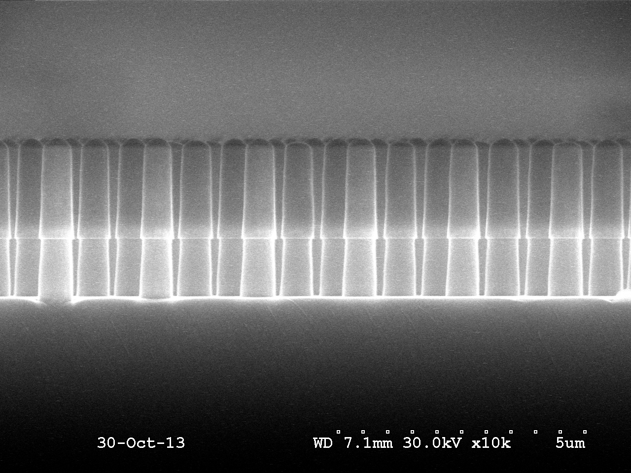
Fig1a_10k_8min_GaNetch.bmp
image/x-ms-bmp (1MB)
Creative Commons: Attribution 4.0
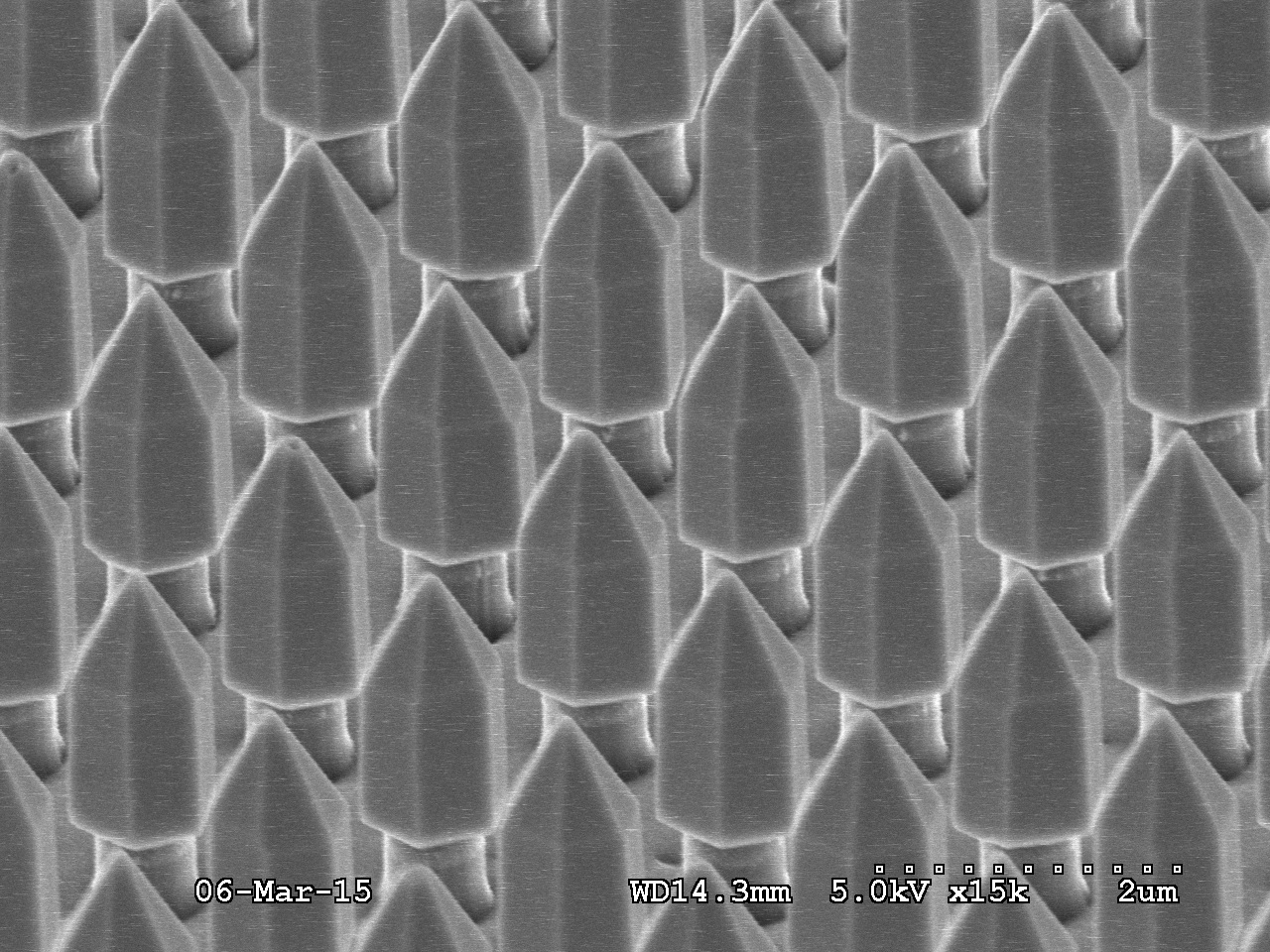
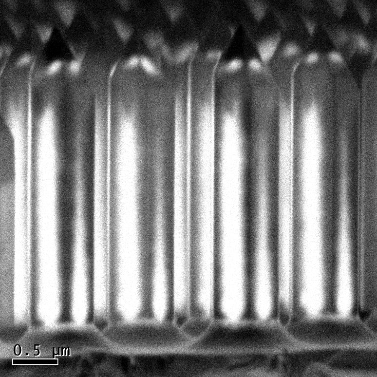
Fig1b_template_cs_10kx.bmp
image/x-ms-bmp (4MB)
Creative Commons: Attribution 4.0
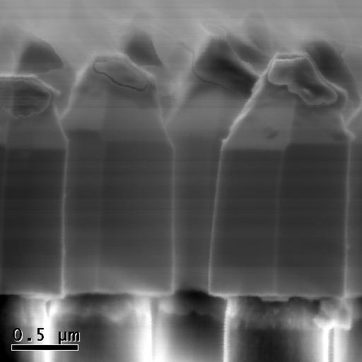
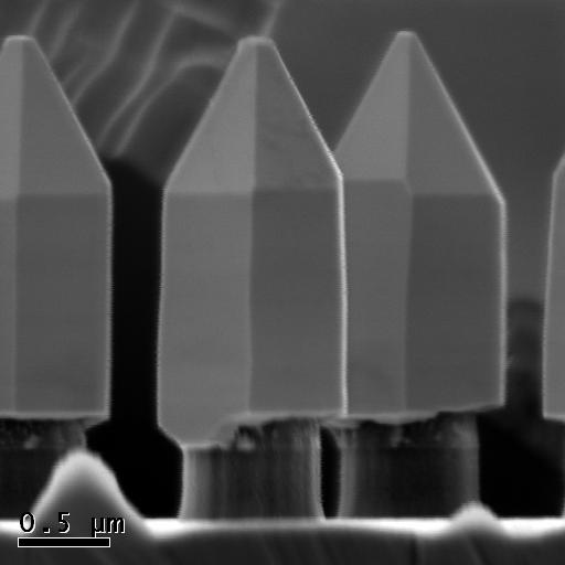
Fig1c_4106_200inj_15x_tilt.bmp
image/x-ms-bmp (1MB)
Creative Commons: Attribution 4.0
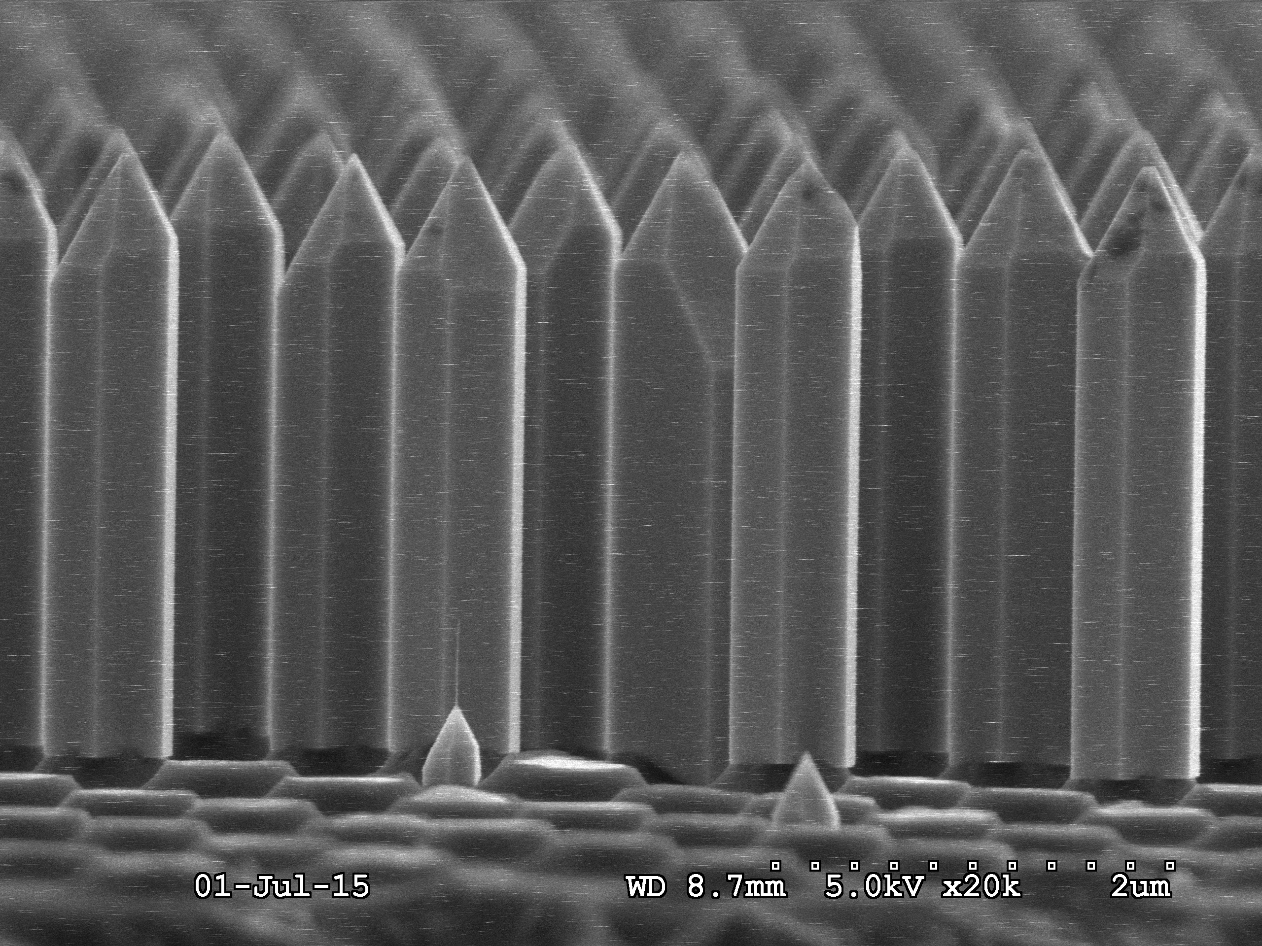
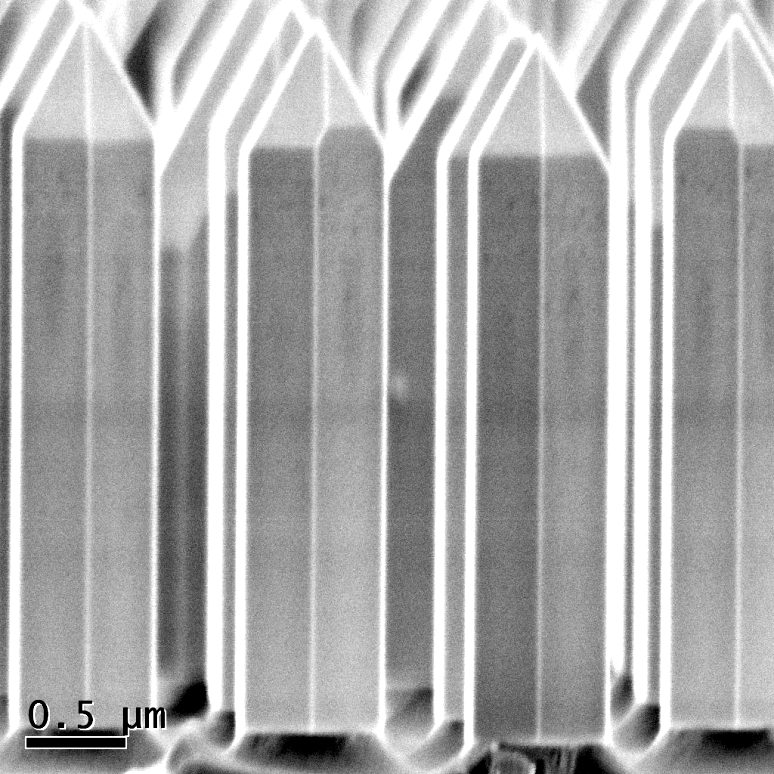
Fig1d_4208_20kx_cs.bmp
image/x-ms-bmp (4MB)
Creative Commons: Attribution 4.0
Fig_2a.csv
text/plain (103kB)
Creative Commons: Attribution 4.0
Fig_2b.csv
text/plain (103kB)
Creative Commons: Attribution 4.0
Fig3a.csv
text/plain (1kB)
Creative Commons: Attribution 4.0
Fig3b.bmp
image/x-ms-bmp (263kB)
Creative Commons: Attribution 4.0
Fig3c.bmp
image/x-ms-bmp (263kB)
Creative Commons: Attribution 4.0
Fig3d.bmp
image/x-ms-bmp (262kB)
Creative Commons: Attribution 4.0
Fig3e.bmp
image/x-ms-bmp (262kB)
Creative Commons: Attribution 4.0
Fig4a.bmp
image/x-ms-bmp (601kB)
Creative Commons: Attribution 4.0
Fig4b.bmp
image/x-ms-bmp (601kB)
Creative Commons: Attribution 4.0
Fig4c.csv
text/plain (5kB)
Creative Commons: Attribution 4.0
Fig4d.bmp
image/x-ms-bmp (601kB)
Creative Commons: Attribution 4.0
Fig4e.bmp
image/x-ms-bmp (601kB)
Creative Commons: Attribution 4.0
Fig4f.csv
text/plain (4kB)
Creative Commons: Attribution 4.0
Fig5a_Raman_y(x,-)ybar.csv
text/plain (43kB)
Creative Commons: Attribution 4.0
Fig5b_Raman_z(x,-)zbar.csv
text/plain (54kB)
Creative Commons: Attribution 4.0
Fig6a_4206-map.txt
text/plain (51MB)
Creative Commons: Attribution 4.0
Fig6b_4208-map-high_res.txt
text/plain (78MB)
Creative Commons: Attribution 4.0
Fig6c_4210-map.txt
text/plain (77MB)
Creative Commons: Attribution 4.0
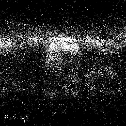
Fig7ab_SE_and … nanorod_C.bmp
image/x-ms-bmp (2MB)
Creative Commons: Attribution 4.0
Fig7c.tif
image/tiff (791kB)
Creative Commons: Attribution 4.0
Fig7d.tif
image/tiff (789kB)
Creative Commons: Attribution 4.0
Creators
Ionut Girgel
University of Bath
Alexander Šatka
Slovak University of Technology in Bratislava
Juraj Priesol
Slovak University of Technology in Bratislava
Pierre-Marie Coulon
University of Bath
Emmanuel Le Boulbar
University of Bath
Tim Batten
Renishaw
Duncan Allsopp
University of Bath
Philip Shields
University of Bath
Contributors
University of Bath
Rights Holder
Documentation
Data collection method:
Secondary electron images were captured using a Hitachi S-4300 scanning electron microscope (SEM). Micro-PL characterization was performed with a He–Cd 325 nm laser excitation source at 0.135 mW power output and a 2400 lines mm−1 diffraction grating in order to obtain a good signal to noise ratio. A 40× magnification, 0.50 numerical aperture (NA) objective produced a spot size of ~0.8 µm, small enough to probe single NRs on a 2 µm pitch. CL maps were obtained on a subset of samples, using an electron beam with a low energy of 2 keV. For the Raman measurements, a high-resolution confocal Raman spectrometer (Renishaw inVia) with a 532 nm laser source, 3000 lines mm−1 grating and 150×, 0.95 NA objective was used. The laser power was 10 mW, the lateral spatial resolution was 0.3 µm and the measurement resolution of the spectral shift was 0.01cm−1. .
Funders
Engineering and Physical Sciences Research Council
https://doi.org/10.13039/501100000266
Lighting the Future
EP/I012591/1
Horizon 2020 Framework Programme
https://doi.org/10.13039/100010661
INREP - Towards Indium free TCOs
641864
Ministry of Education, Science, Research and Sport of the Slovak Republic
https://doi.org/10.13039/501100006109
Analysis and characterization of advanced power electronic devices supported by 2/3D electrothermal modeling and simulation
1/0491/15
Publication details
Publication date: 27 March 2018
by: University of Bath
Version: 1
DOI: https://doi.org/10.15125/BATH-00207
URL for this record: https://researchdata.bath.ac.uk/207
Related papers and books
Gîrgel, I., Šatka, A., Priesol, J., Coulon, P.-M., Le Boulbar, E. D., Batten, T., Allsopp, D. W. E., and Shields, P. A., 2018. Optical characterization of magnesium incorporation in p-GaN layers for core–shell nanorod light-emitting diodes. Journal of Physics D: Applied Physics, 51(15), 155103. Available from: https://doi.org/10.1088/1361-6463/aab16b.
Contact information
Please contact the Research Data Service in the first instance for all matters concerning this item.
Faculty of Engineering & Design
Electronic & Electrical Engineering
