Dataset for Investigation of InGaN facet-dependent non-polar growth rates and composition for core-shell LEDs
Core-shell InGaN/GaN structures are attractive as light emitters due to their potential for emission on large non-polar surface area and flexibility in achieving different emission wavelengths with changing growth parameters. The morphology and dimensional dependence of InGaN layers with growth parameters were investigated by scanning electron microscopy (SEM) and transmission electron microscopy (TEM). Growth rates for InGaN under different growth parameters for the non-polar a- and m-planes were determined.
This dataset contains the results of scanning electron microscopy (SEM) and transmission electron microscopy (TEM) measurements carried out on core-shell nanostructures. The samples are highly regular arrays of GaN plasma etched cores onto which thick InGaN layers were grown using different metal organic vapour phase epitaxy (MOVPE) growth parameters.
Three different InGaN growth conditions were considered with the following parameters: 750°C at 300 mbar, 700°C at 300 mbar and 750°C at 100 mbar. Statistical growth rates were determined on the non-polar crystal planes from measurements of increase in diameter using SEM images. TEM analysis was carried out on a single nanorod for greater detail.
Cite this dataset as:
Girgel, I.,
Le Boulbar, E.,
Coulon, P.,
Sahonta, S.,
Allsopp, D.,
Humphreys, C.,
Shields, P.,
2016.
Dataset for Investigation of InGaN facet-dependent non-polar growth rates and composition for core-shell LEDs.
Bath: University of Bath Research Data Archive.
Available from: https://doi.org/10.15125/BATH-00172.
Export
Data
3872_gan_6x_25s.bmp
image/x-ms-bmp (4MB)
Creative Commons: Attribution 4.0
SEM image of GaN regrowth sample
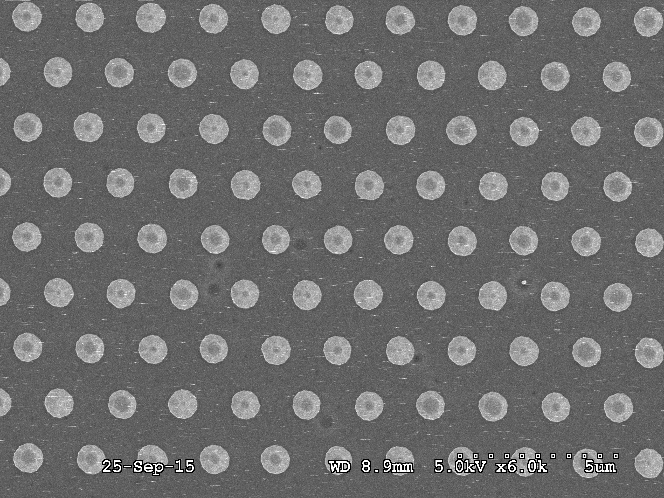
3873_ingan_6x__2nd_25s.bmp
image/x-ms-bmp (4MB)
Creative Commons: Attribution 4.0
SEM image of InGaN sample grown at 750 C and 300 mbar.
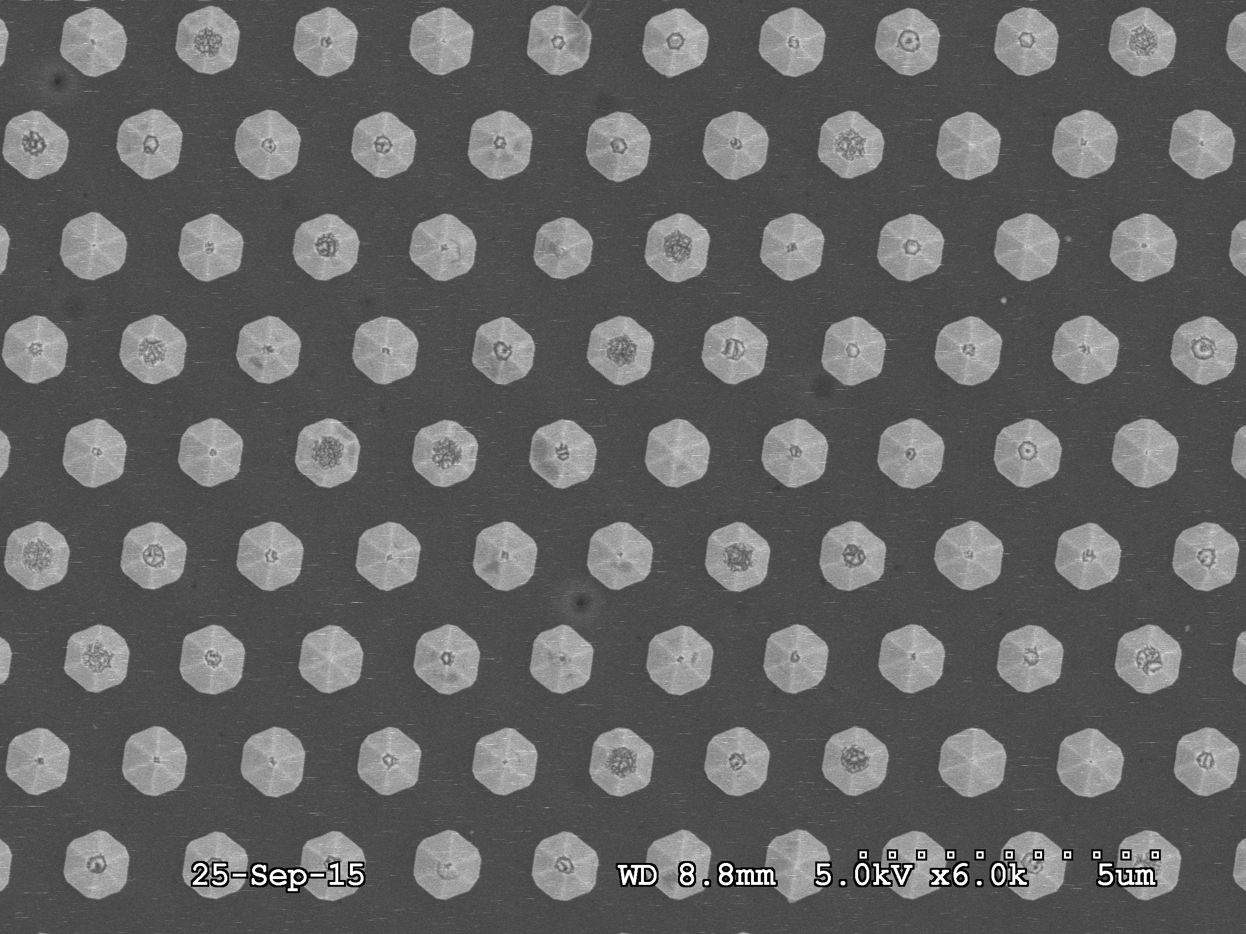
3874_ingan_6x_25s.bmp
image/x-ms-bmp (4MB)
Creative Commons: Attribution 4.0
SEM image of InGaN sample grown at 700 C and 300 mbar.
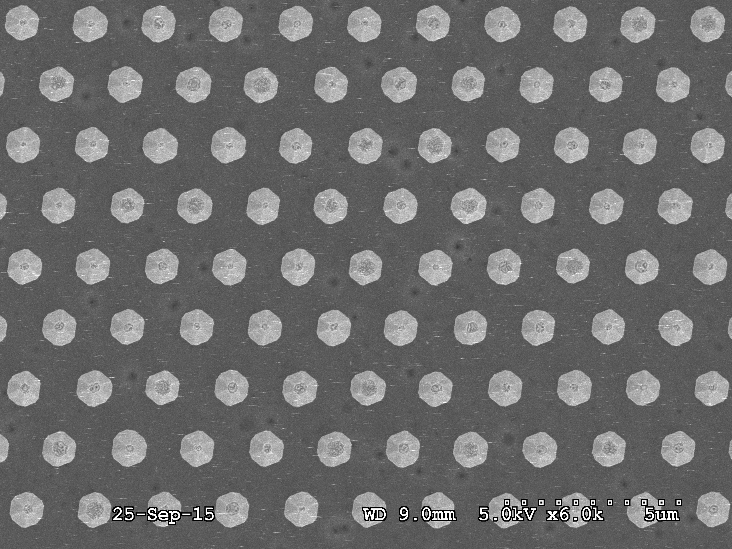
3875_gan_6x_25s.bmp
image/x-ms-bmp (4MB)
Creative Commons: Attribution 4.0
SEM image of InGaN sample grown at 750 C and 100 mbar.
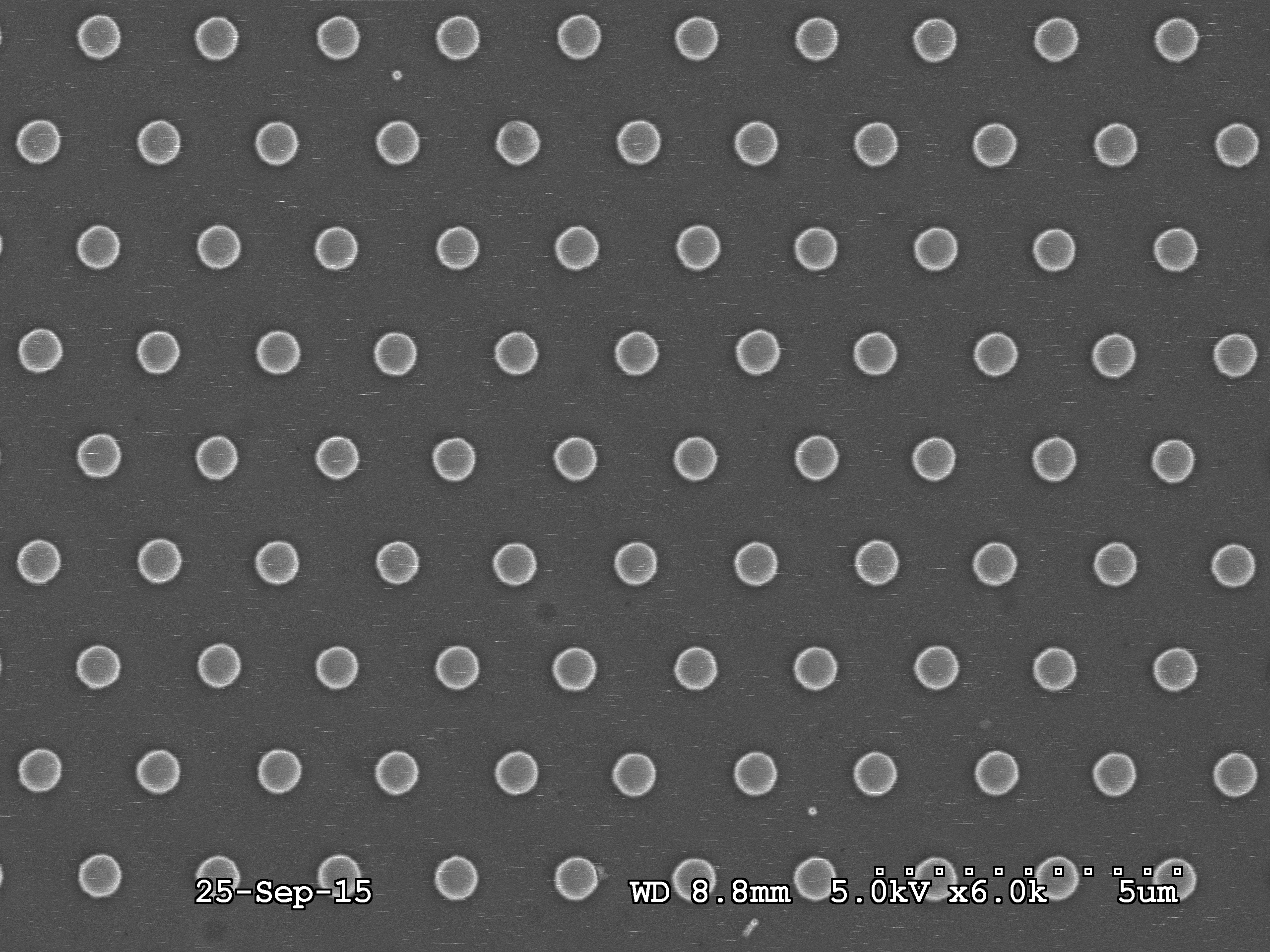
template_6x_25s.bmp
image/x-ms-bmp (4MB)
Creative Commons: Attribution 4.0
SEM image of plasma etched GaN template.
161424.tif
image/tiff (2MB)
Creative Commons: Attribution 4.0
TEM image of planar cross section of InGaN sample grown at 700 C and 300 mbar.
3872_130thresh_2filter.xlsx
application/vnd.openxmlformats-officedocument.spreadsheetml.sheet (12kB)
Creative Commons: Attribution 4.0
3873_130thresh_2filter.xlsx
application/vnd.openxmlformats-officedocument.spreadsheetml.sheet (13kB)
Creative Commons: Attribution 4.0
3874_130thresh_2filter.xlsx
application/vnd.openxmlformats-officedocument.spreadsheetml.sheet (13kB)
Creative Commons: Attribution 4.0
3875_130thresh_2filter.xlsx
application/vnd.openxmlformats-officedocument.spreadsheetml.sheet (13kB)
Creative Commons: Attribution 4.0
template_130thresh_3filter.xlsx
application/vnd.openxmlformats-officedocument.spreadsheetml.sheet (13kB)
Creative Commons: Attribution 4.0
centraliz_values … rates.xlsx
application/vnd.openxmlformats-officedocument.spreadsheetml.sheet (9kB)
Creative Commons: Attribution 4.0
tem_vis_assist_r2.xlsx
application/vnd.openxmlformats-officedocument.spreadsheetml.sheet (10kB)
Creative Commons: Attribution 4.0
tem_m-a_ratio.xlsx
application/vnd.openxmlformats-officedocument.spreadsheetml.sheet (10kB)
Creative Commons: Attribution 4.0
All data are available under a Creative Commons Attribution 4.0 (CC-BY) licence.
Creators
Ionut Girgel
University of Bath
Emmanuel Le Boulbar
University of Bath
Pierre-Marie Coulon
University of Bath
Suman-Lata Sahonta
University of Cambridge
Duncan Allsopp
Supervisor
University of Bath
Colin J. Humphreys
Supervisor
University of Cambridge
Philip Shields
University of Bath
Contributors
University of Bath
Rights Holder
University of Cambridge
Rights Holder
Documentation
Data collection method:
Secondary electron images were captured using a Hitachi S-4300 scanning electron microscope (SEM). An accelerating voltage of 5 kV was used to collect the images at constant magnification of 6000x from 5 samples, corresponding to a plasma etched GaN template, a GaN regrowth and three InGaN growths. Approximately 80 individual nanorods and their diameters are identified in each image. Each image was processed by binary threshold and low pass filter in Vision Assistant 2011 to obtain individual nanorod dimensions as detailed in the *.xls files. Each xls file contains separate columns for object number, calibrated area, equivalent disk diameter and image area. Statistical distributions of diameters were determined. A planar cross section was prepared for TEM measurements using a focused ion beam to thin down the cross section to 100-200 nm thickness. A Tecnai Osiris scanning-TEM (STEM) operating at 200 kV was used to obtain a high-angle annular dark-field (HAADF) Z-contrast image. The same image processing as in the SEM images was used to determine diameters and InGaN shell thickness.
Technical details and requirements:
Hitachi S-4300 scanning electron microscope (SEM) Focused Ion Beam (FIB) etching and platinum deposition Tecnai Osiris scanning transmission electron microscope (STEM)
Funders
Seventh Framework Programme
https://doi.org/10.13039/501100004963
SMASH: Smart Nanostructured Semiconductors for Energy-Saving Light Solutions
228999
Engineering and Physical Sciences Research Council
https://doi.org/10.13039/501100000266
Lighting the Future
EP/I012591/1
Publication details
Publication date: 27 January 2016
by: University of Bath
Version: 1
DOI: https://doi.org/10.15125/BATH-00172
URL for this record: https://researchdata.bath.ac.uk/172
Related papers and books
Gîrgel, I., Edwards, P. R., Le Boulbar, E., Coulon, P.-M., Sahonta, S.-L., Allsopp, D. W. E., Martin, R. W., Humphreys, C. J., and Shields, P. A., 2016. Investigation of indium gallium nitride facet-dependent nonpolar growth rates and composition for core–shell light-emitting diodes. Journal of Nanophotonics, 10(1), 016010. Available from: https://doi.org/10.1117/1.jnp.10.016010.
Related datasets and code
Edwards, P., and Martin, R., 2016. Cathodoluminescence imaging and spectroscopy of InGaN/GaN core-shell nanostructures. University of Strathclyde. Available from: https://doi.org/10.15129/53C3BD47-91D8-40CE-A6AC-98E46B893588.
Contact information
Please contact the Research Data Service in the first instance for all matters concerning this item.
Contact person: Ionut Girgel
Faculty of Engineering & Design
Electronic & Electrical Engineering
